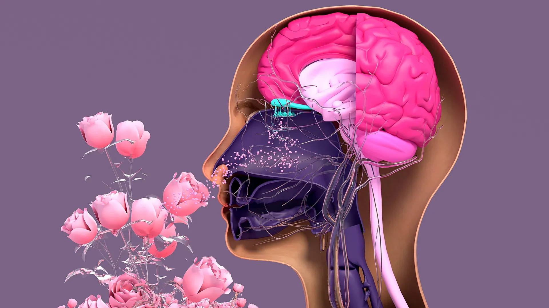
In a significant breakthrough for regenerative medicine, researchers at Tufts University School of Medicine and the Graduate School of Biomedical Sciences (GSBS) have developed a novel three-dimensional model to study how nerve tissue in the nose repairs itself. This study, recently published in Cell Reports Methods, has overturned long-held assumptions regarding stem cell activity in the olfactory system, revealing that a cell type previously dismissed as dormant plays a foundational role in maintaining our sense of smell.
Unlike the neurons within the central nervous system, which are notoriously difficult to regenerate, the sensory neurons lining the human nasal cavity possess a remarkable, lifelong capacity for renewal. This continuous turnover is essential because these cells are in direct, constant contact with the external environment, leaving them perpetually vulnerable to inhaled toxins, pathogens, and environmental pollutants. However, when this regenerative engine falters—due to chronic viral infections, such as those caused by COVID-19, long-term exposure to hazardous substances, or the natural degradation that accompanies aging—individuals can suffer from partial or complete anosmia, or the loss of smell.
Unmasking the Role of Basal Cells
The research team, led by senior author Brian Lin, a research assistant professor in the Department of Developmental, Molecular and Chemical Biology at Tufts, sought to create a more accessible way to investigate why this regenerative process eventually declines. To achieve this, the team engineered an easy-to-create, three-dimensional olfactory tissue mouse model, known as an organoid. This miniature, lab-grown version of nasal tissue allows scientists to observe the microscopic choreography of cellular regeneration in real time.
Within the nasal epithelium, two primary types of stem cells are responsible for the renewal of smell-sensing neurons: horizontal basal cells (HBCs) and globose basal cells (GBCs). Traditionally, the scientific community viewed GBCs as the primary workhorses of regeneration, while HBCs were largely characterized as a dormant reserve, activated only during extreme injury.
However, the Tufts team’s findings suggest a more nuanced and interdependent relationship. Using the new organoid model, the researchers identified a specific subpopulation of HBCs, defined by their expression of the protein KRT5, that actively contribute to the ongoing generation of new olfactory neurons.
"Our research suggests that these two stem cells may be interdependent," explains Dr. Lin. "One type that we thought was largely dormant—HBCs—may actually play a crucial role in supporting the production of new neurons and the repair of damaged tissue."
To validate this discovery, the researchers selectively depleted these KRT5-producing HBCs from the organoid cultures. The result was a significant impairment in the generation of new neurons, confirming that these cells are not merely bystanders or dormant reserves, but essential, active participants in the regenerative ecosystem. The study suggests that the interplay between HBCs and GBCs is a finely tuned mechanism, and when that communication is disrupted, the capacity of the nose to replace damaged sensory neurons is severely compromised.
The Impact of Aging on Sensory Regeneration
Beyond identifying the critical role of HBCs, the research team investigated how the regenerative capacity of these cells changes over the course of a lifespan. By utilizing the 3D organoid model to grow cells taken from mice of varying ages, the researchers were able to quantify the decline in neurogenesis that occurs with maturity.
"We found a decline in the ability of the older mice cells to generate new neurons," Lin notes. "We think this is due to a decrease in the GBC population as we age, but we need to do more work to test this hypothesis and, if so, develop ways to rejuvenate them."
This finding provides a potential biological explanation for why the sense of smell often diminishes in older populations. If the population of GBCs naturally dwindles over time, the olfactory system may struggle to maintain the pace of neuron turnover required to keep the sense of smell sharp. Understanding this mechanism is the first step toward potential future therapies aimed at "rejuvenating" these stem cell populations to restore sensory function.
Advancing Research Through Accessible Models
A major priority for the research team was ensuring that their new 3D model would be accessible to a wide range of laboratories. Lead author Juliana Gutschow Gameiro, a visiting Ph.D. student at GSBS from the State University of Londrina in Brazil, focused her efforts on developing a protocol that is straightforward and requires minimal specialized equipment or funding.
Dr. Lin emphasizes the importance of this accessibility, especially given the surge in interest regarding olfactory health. "Because loss of smell is associated with COVID-19, as well as with Parkinson’s disease and other conditions, a much larger number of researchers from a variety of different fields have begun researching olfactory epithelial cells in the last few years," he says.
By creating an easy-to-use organoid, the team hopes to lower the barrier to entry for scientists who may not be stem cell experts but are nonetheless invested in understanding how olfactory neurons fail and how they might be repaired. This democratization of research tools could significantly accelerate the pace of discovery in the field of sensory biology.
The Path Toward Human Organoids
The ultimate ambition for the Tufts team is to translate their success with the mouse-tissue model into a human organoid. Such a model would serve as a powerful, high-throughput platform for screening pharmaceutical drugs, potentially offering new treatments for patients suffering from long-term olfactory loss.
Organoids have already proven their value in clinical research, with models currently used to study the biology of lungs, kidneys, and other vital organs. These systems offer a significant advantage over animal models or traditional two-dimensional cell cultures, providing a more accurate physiological representation of human tissue while reducing the time and cost associated with pre-clinical drug development.
However, moving from a mouse model to a human one presents distinct logistical challenges. Unlike the mouse model, where stem cells can be clearly defined and isolated, collecting pure olfactory tissue from humans is complex. The standard procedure—anesthetizing a patient and using a brush-like device to collect cells from the deep nasal cavity—often results in a mixture of both respiratory and olfactory stem cells. Distinguishing between these two cell types remains a hurdle for researchers.
The next phase of the team’s research will focus on developing a refined, inexpensive technique to effectively separate human olfactory stem cells from respiratory cells and coax them into thriving within a laboratory environment. If successful, this would unlock the ability to model human olfactory disease with unprecedented precision.
As the scientific community continues to grapple with the long-term sensory consequences of global health events and age-related decline, the work being done at Tufts provides both a new framework for study and a glimmer of hope for future therapies. By shifting the focus toward the complex, interdependent nature of nasal stem cells, researchers are moving closer to understanding the cellular blueprints required to restore one of the most vital—and often overlooked—human senses.