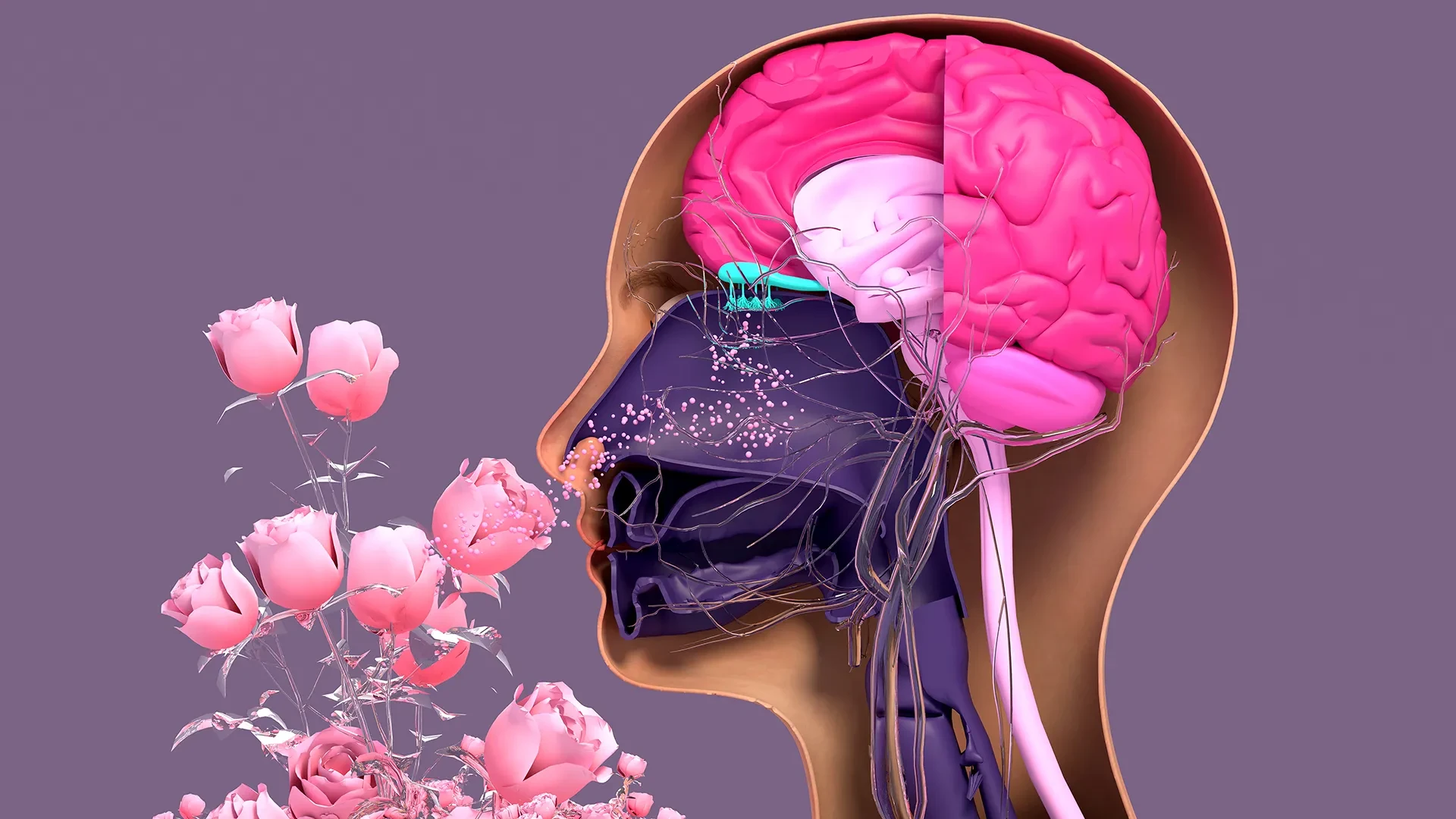
In a significant stride toward understanding how the human body repairs its sensory systems, researchers at Tufts University School of Medicine and the Graduate School of Biomedical Sciences (GSBS) have unveiled a new, three-dimensional model for studying the regeneration of nerve tissue in the nose. This breakthrough, recently detailed in the journal Cell Reports Methods, has led to the surprising discovery that a specific type of stem cell, long assumed to be largely dormant, plays a far more active role in preserving the sense of smell than previously acknowledged by the scientific community.
Unlike the central nervous system, which possesses a limited capacity for repair, the sensory neurons within the human nasal cavity exhibit a remarkable, lifelong ability to regenerate. This continuous renewal is essential, given that these neurons are in constant contact with the outside world, frequently exposed to environmental toxins, pathogens, and physical trauma. However, this regenerative capacity is not invincible. Viral infections—most notably SARS-CoV-2—prolonged exposure to hazardous chemicals, and the natural physiological decline associated with aging can all impair the function of these cells. When these regenerative processes falter, the result is often a partial or complete loss of smell, a condition that can have profound impacts on a patient’s quality of life, nutritional health, and psychological well-being.
To better investigate the mechanics behind this continuous cellular turnover and the reasons for its eventual failure, the Tufts research team developed a novel, 3D olfactory tissue mouse model, known as an organoid. These organoids serve as miniature, simplified versions of complex biological systems, allowing scientists to observe cellular interactions in a controlled laboratory environment. By utilizing this model, the team successfully mapped the complex communication network between two primary types of stem cells in the nose: horizontal basal cells (HBCs) and globose basal cells (GBCs).
Historically, the scientific consensus categorized HBCs as a relatively quiescent, or "dormant," cell population that only engaged in tissue repair during instances of severe, widespread injury. However, the study conducted by the Tufts team challenges this paradigm. "Our research suggests that these two stem cells may be interdependent," explains Brian Lin, a research assistant professor in the Department of Developmental, Molecular and Chemical Biology and the senior author of the study. "One type that we thought was largely dormant—HBCs—may actually play a crucial role in supporting the production of new neurons and the repair of damaged tissue."
Through their organoid model, the researchers identified a distinct subpopulation of HBCs, characterized by the production of the protein KRT5. These cells appear to be vital, active participants in the generation of new olfactory neurons. When the team experimentally depleted these KRT5-producing HBCs from the organoid cultures, they observed a significant impairment in the generation of new sensory neurons. This finding provides compelling evidence that these cells are not merely a reserve force waiting for catastrophe, but are instead fundamental players in the daily maintenance and regenerative health of the olfactory system.
The study also shed light on why the sense of smell often diminishes as organisms age. By cultivating cells from mice of different ages within the 3D model, the researchers were able to compare regenerative efficiency across the lifespan. "We found a decline in the ability of the older mice cells to generate new neurons," Lin noted. "We think this is due to a decrease in the GBC population as we age, but we need to do more work to test this hypothesis and, if so, develop ways to rejuvenate them."
An Accessible Model for Global Research
A primary objective for the team, led by primary author Juliana Gutschow Gameiro—a former visiting Ph.D. student at GSBS from the State University of Londrina in Brazil—was the creation of a model that is both accessible and cost-effective. Developing sophisticated organoids can often be an expensive and technically demanding endeavor, which frequently limits research to well-funded, large-scale laboratories.
Given the global surge in interest surrounding olfactory health, particularly in the wake of the COVID-19 pandemic, the demand for accessible research tools has never been higher. "Because loss of smell is associated with COVID-19, as well as with Parkinson’s disease and other conditions, a much larger number of researchers from a variety of different fields have begun researching olfactory epithelial cells in the last few years," Lin says. "We wanted to develop an easy-to-use model so that non-stem cell biologists and those working in labs with limited resources could use it to better understand how olfactory neurons regenerate and what happens that causes that process to diminish or fail completely."
By lowering the barrier to entry for studying olfactory epithelial cells, the team hopes to catalyze a broader, more collaborative effort to solve the mysteries of sensory loss. The model’s design is intended to provide a robust, repeatable platform for investigating the molecular pathways of neural regeneration without the need for specialized equipment that might otherwise be unavailable in smaller research settings.
Paving the Way for Human Organoid Therapy
While the mouse-tissue model represents a substantial leap forward, the ultimate goal of the Tufts researchers is the development of a human organoid. Such a model would be an invaluable asset for drug screening and the development of clinical therapies for patients suffering from anosmia—the total loss of smell—or hyposmia, the diminished ability to detect odors.
The transition from a mouse model to a human one, however, presents significant biological and logistical challenges. One of the primary hurdles is the difficulty of obtaining pure olfactory tissue from human subjects. The current standard procedure involves inserting a brush—similar to a swab used for COVID-19 testing—deep into the nasal cavity while the patient is anesthetized. During this collection, the olfactory stem cells are often gathered alongside respiratory stem cells, which are structurally similar and difficult to isolate from one another.
"It’s challenging to get pure olfactory tissue from humans," Lin notes, explaining that the contamination of the sample with respiratory cells can complicate the growth and analysis of the olfactory stem cells in a laboratory setting.
Despite these obstacles, the potential benefits of perfecting a human organoid system are immense. Compared to traditional methods of drug testing, which rely on whole animals or limited two-dimensional human cell cultures, organoids provide a more accurate representation of the human physiological environment. This allows for faster, less expensive, and more effective pre-clinical research. Organoids have already been successfully pioneered for other human organs, including the lungs and kidneys, providing a roadmap that the team hopes to follow for olfactory tissue.
The team’s current research trajectory is focused on overcoming the bottleneck of cell separation. By developing a simple, cost-effective technique to reliably isolate human olfactory stem cells and encourage them to proliferate within the laboratory, the researchers aim to bridge the gap between basic cellular biology and clinical application. As they continue to refine their models, the focus remains on transforming these biological insights into tangible medical interventions. With the global population aging and the long-term impacts of respiratory viruses remaining a public health concern, the ability to stimulate the regeneration of the sense of smell could significantly improve the quality of life for millions of people worldwide. The work at Tufts University serves as a foundational step toward that future, proving that even the most "dormant" cells may hold the key to restoring one of our most vital senses.