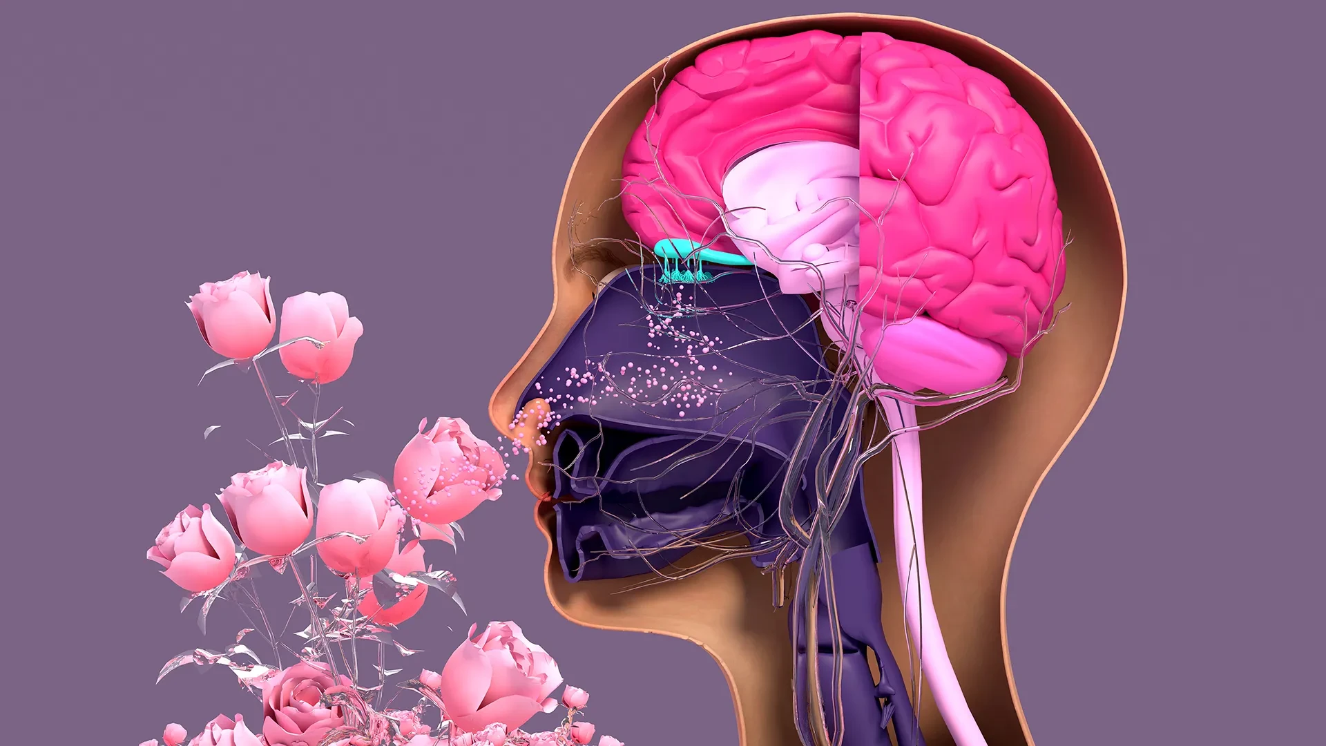
In a significant advancement for regenerative medicine and sensory biology, researchers at Tufts University School of Medicine and the Graduate School of Biomedical Sciences (GSBS) have unlocked new insights into how the nose repairs itself. By developing a novel, three-dimensional model to study nasal tissue, the team has discovered that a specific type of stem cell—previously dismissed as dormant—actually serves as a vital architect in the preservation and repair of our sense of smell.
The research, published in the journal Cell Reports Methods, provides a sophisticated yet accessible framework for studying olfactory neurons. This breakthrough comes at a critical time, as the medical community continues to grapple with the long-term sensory consequences of viral infections like COVID-19, the impact of environmental toxins, and the natural decline of sensory function associated with aging.
Understanding the Resilience of the Olfactory System
Unlike the delicate and often permanent damage seen in the central nervous system, the sensory neurons within the nasal cavity possess a unique and remarkable capacity for regeneration. Throughout a human lifetime, these neurons are constantly exposed to the external environment, including pathogens, pollutants, and irritants. To survive this onslaught, the olfactory epithelium—the specialized tissue responsible for detecting odors—must undergo a continuous process of renewal, replacing dead or damaged neurons with new ones generated from resident stem cell populations.
However, this regenerative capacity is not invincible. Factors such as severe viral infections, chronic exposure to hazardous chemicals, and the cumulative effects of aging can overwhelm the nose’s self-repair mechanisms. When these systems falter, the result is often a partial or complete loss of smell, known clinically as anosmia or hyposmia. Understanding the cellular "why" behind this decline has long been a goal for researchers, but progress has been hampered by the difficulty of modeling this complex environment in a laboratory setting.
The Power of the Organoid
To overcome these barriers, the Tufts research team, led by senior author Brian Lin, a research assistant professor in the Department of Developmental, Molecular and Chemical Biology, devised a three-dimensional olfactory tissue model, or "organoid." These miniature, lab-grown versions of organ tissue allow scientists to observe cellular interactions in a controlled environment that mimics the complexity of a living organism.
The study utilized this mouse-derived organoid model to map the communication between two primary types of stem cells: horizontal basal cells (HBCs) and globose basal cells (GBCs). Traditionally, the scientific consensus held that GBCs were the primary workhorses of neurogenesis, actively dividing to create new neurons, while HBCs were largely considered a dormant reserve, only activated in the event of extreme tissue injury.
The new findings, however, challenge this paradigm. "Our research suggests that these two stem cells may be interdependent," Lin explains. "One type that we thought was largely dormant—HBCs—may actually play a crucial role in supporting the production of new neurons and the repair of damaged tissue."
By focusing on a specific subpopulation of HBCs identified by their expression of the protein KRT5, the team observed that these cells are far more active than previously documented. In the organoid environment, these KRT5-positive HBCs were instrumental in the initial formation and structural integrity of the tissue. When the researchers selectively depleted these HBCs from the cultures, the generation of new olfactory neurons plummeted, confirming that these cells are essential, active players in the regenerative cycle rather than mere bystanders waiting for a crisis.
The Challenge of Aging
The research team also investigated how this regenerative process shifts over time. By cultivating cells from mice of varying ages, the team observed a distinct decline in the capacity of older cells to generate new neurons. Lin and his colleagues hypothesize that this decline is linked to a reduction in the GBC population as the organism ages.
"We found a decline in the ability of the older mice cells to generate new neurons," Lin says. "We think this is due to a decrease in the GBC population as we age, but we need to do more work to test this hypothesis and, if so, develop ways to rejuvenate them." This line of inquiry opens doors for future therapeutic interventions that could potentially target and "recharge" these stem cell populations, offering hope for treating age-related olfactory loss.
A Democratized Tool for Research
A central priority for the project was to ensure the model could be widely adopted. The study was spearheaded by lead author Juliana Gutschow Gameiro, a visiting Ph.D. student from the State University of Londrina in Brazil. Under Lin’s mentorship, Gameiro focused on developing a protocol that would be easy to replicate in laboratories, even those with limited funding or specialized equipment.
This accessibility is essential because of the growing interest in olfactory research. As the prevalence of smell loss has risen in the wake of the COVID-19 pandemic—and given the well-established link between olfactory dysfunction and neurodegenerative conditions like Parkinson’s disease—the field has seen an influx of researchers from various disciplines.
"We wanted to develop an easy-to-use model so that non-stem cell biologists and those working in labs with limited resources could use it to better understand how olfactory neurons regenerate and what happens that causes that process to diminish or fail completely," Lin notes. By lowering the barrier to entry, the team hopes to accelerate the pace of discovery across the global scientific community.
Moving Toward Human Applications
While the mouse-tissue organoid is a significant success, it is viewed as a stepping stone toward the ultimate objective: the creation of a human olfactory organoid. Such a tool would be invaluable for high-throughput drug screening, allowing researchers to test potential treatments for olfactory loss in a human-relevant model before proceeding to clinical trials.
Organoids have already transformed research for other organs, including the lungs and kidneys, providing a more effective and ethical alternative to animal testing or traditional two-dimensional cell cultures. However, the nose presents a unique hurdle. Extracting pure olfactory tissue from humans is technically demanding; it requires an invasive procedure where a brush is inserted deep into the nasal cavity under anesthesia. Currently, the samples collected in this manner contain a mixture of respiratory and olfactory stem cells that are notoriously difficult to separate.
The next phase of the team’s research will focus on overcoming this biological bottleneck. Their challenge is to refine a simple, cost-effective technique that can isolate human olfactory stem cells from these mixed samples and successfully coax them into healthy, self-sustaining organoids.
If the team can successfully adapt their mouse-model success to human tissue, it would represent a transformative shift in how we treat sensory loss. By unlocking the regenerative secrets of the nose, researchers are not only gaining a better understanding of how we perceive the world around us, but they are also building the foundation for therapies that could one day restore the sense of smell to millions of people worldwide. As the project continues to evolve, the combination of accessible methodology and rigorous biological discovery promises to keep Tufts at the forefront of olfactory research.