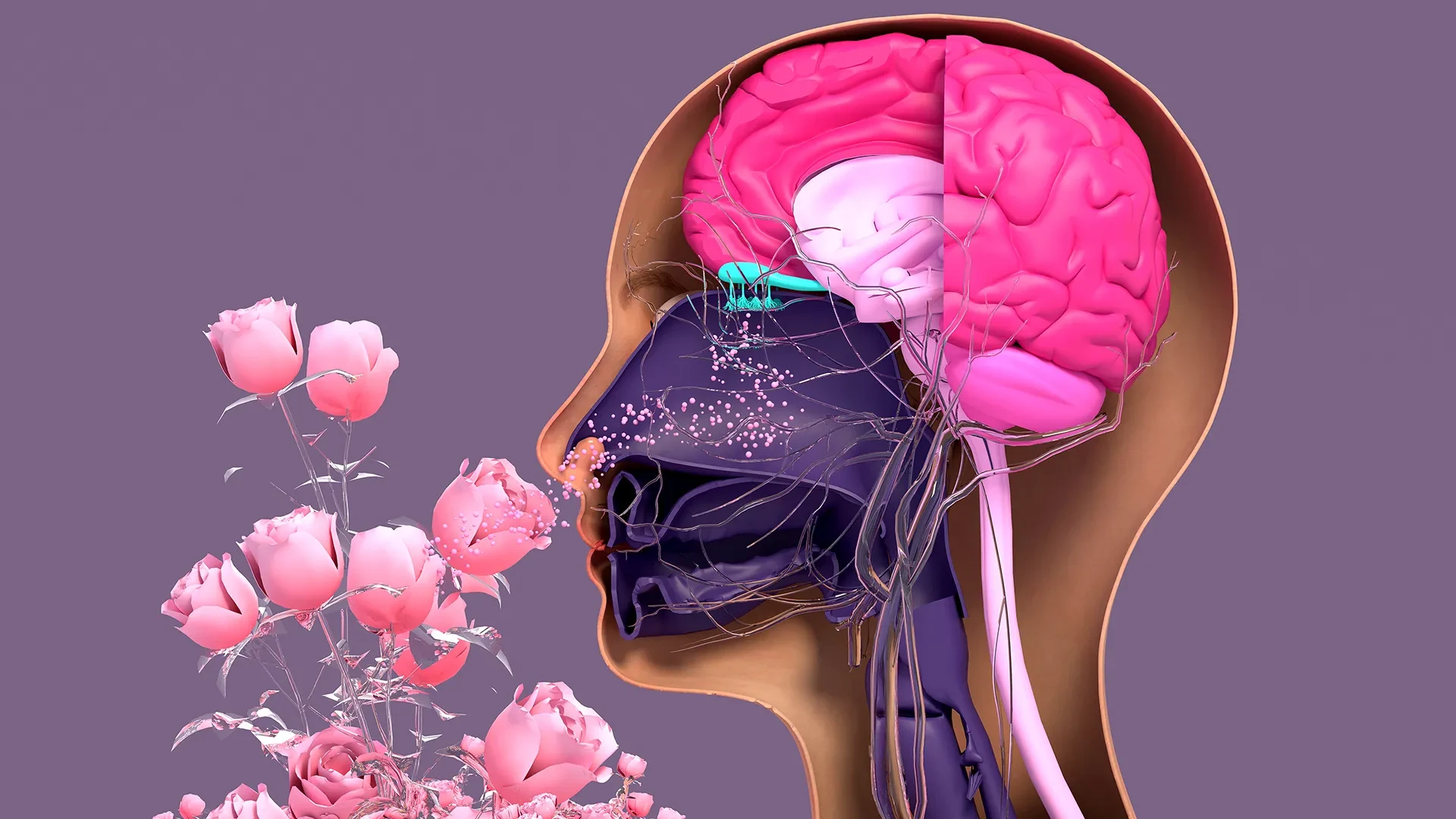
In a significant breakthrough for sensory biology, researchers at Tufts University School of Medicine and the Graduate School of Biomedical Sciences (GSBS) have unveiled a new three-dimensional model that sheds light on the complex process of olfactory regeneration. The study, published in Cell Reports Methods, reveals that certain stem cells previously thought to be largely dormant in the nose may actually serve as vital architects in the maintenance and repair of the sense of smell.
Unlike the central nervous system, where damage is often permanent, the sensory neurons within the human nasal cavity possess a remarkable, lifelong capacity for regeneration. This continuous turnover allows the body to replace neurons exposed to the harsh, ever-changing outside environment. However, this regenerative engine is not immune to failure. Viral infections—most notably COVID-19—as well as exposure to environmental toxins and the natural processes of aging, can severely impair the function or replication of these cells. When this mechanism falters, individuals may experience a partial or total loss of their sense of smell, a condition that can profoundly impact quality of life, nutritional intake, and emotional well-being.
To better understand why this biological process declines, the research team, led by senior author Brian Lin, a research assistant professor in the Department of Developmental, Molecular and Chemical Biology, developed a novel, accessible 3D olfactory tissue mouse model. By utilizing these "organoids"—miniature, simplified versions of organs grown in the laboratory—scientists can now observe the intricate dance of cellular communication that facilitates the birth of new smell-sensing neurons.
Decoding the Cellular Dialogue
The study focuses on the interaction between two primary types of stem cells found in the nasal epithelium: horizontal basal cells (HBCs) and globose basal cells (GBCs). Traditionally, the scientific community categorized HBCs as a quiescent, or dormant, population, only recruited during times of extreme tissue injury. GBCs, by contrast, were viewed as the primary, active workhorses of daily neuronal turnover.
However, the findings from the Tufts team suggest a more nuanced and interdependent relationship. Through the use of their 3D organoid model, the researchers identified a specific subpopulation of HBCs, characterized by the production of the protein KRT5, which plays a much more active role in tissue maintenance than previously understood.
"Our research suggests that these two stem cells may be interdependent," says Lin. "One type that we thought was largely dormant—HBCs—may actually play a crucial role in supporting the production of new neurons and the repair of damaged tissue."
To confirm this, the team conducted depletion experiments within their organoid cultures. When the KRT5-expressing HBCs were selectively removed, the researchers observed a significant impairment in the generation of new olfactory neurons. This clear cause-and-effect relationship confirms that HBCs are not merely reserves waiting for a catastrophe; they are essential, ongoing participants in the regenerative process.
Furthermore, the team investigated the impact of age on these cellular dynamics. By comparing cells derived from mice of different ages, the researchers noted a distinct decline in the regenerative capacity of cells taken from older subjects. While the exact mechanism is still under investigation, Lin and his colleagues hypothesize that this decline is linked to a reduction in the GBC population as the organism ages. "We think this is due to a decrease in the GBC population as we age," Lin explains, "but we need to do more work to test this hypothesis and, if so, develop ways to rejuvenate them."
A Democratized Tool for Scientific Discovery
A pivotal aspect of the study was the focus on accessibility. The lead author of the study, Juliana Gutschow Gameiro—a former Ph.D. student who visited the GSBS from the State University of Londrina in Brazil—prioritized the creation of a model that could be easily replicated by laboratories with limited budgets or sophisticated equipment.
This focus on accessibility is intentional and timely. As the global scientific community grapples with the long-term sensory effects of the COVID-19 pandemic, interest in olfactory biology has surged. Researchers from a wide range of disciplines, many of whom may not specialize in stem cell biology, are now racing to understand the pathology of smell loss.
"Because loss of smell is associated with COVID-19, as well as with Parkinson’s disease and other conditions, a much larger number of researchers from a variety of different fields have begun researching olfactory epithelial cells in the last few years," says Lin. "We wanted to develop an easy-to-use model so that non-stem cell biologists and those working in labs with limited resources could use it to better understand how olfactory neurons regenerate and what happens that causes that process to diminish or fail completely."
The development of this model provides a scalable platform for studying these cells in a controlled environment, moving beyond the limitations of whole-animal studies or static cell cultures.
The Road Toward Human Organoids
The ultimate ambition of the Tufts team is to translate this mouse-based success into a human organoid model. Such a breakthrough would be a transformative development for clinical research, offering a powerful tool for high-throughput drug screening. If researchers can grow human olfactory tissue in the lab, they could potentially test a library of compounds to identify those capable of stimulating regeneration in patients suffering from persistent anosmia, or the loss of smell, due to disease or injury.
Organoids have already revolutionized the study of other organs, including the lungs and kidneys, by providing a bridge between simple cell cultures and complex human clinical trials. However, human olfactory tissue presents a unique set of technical hurdles.
"It’s challenging to get pure olfactory tissue from humans," Lin notes. The standard collection method involves a procedure similar to a nasal swab used for COVID-19 testing, where a brush is inserted deep into the nasal cavity. While effective for collecting samples, this method harvests a mixture of respiratory and olfactory stem cells. Distinguishing and separating these two cell types remains a significant challenge, as they are often harvested together and require specific conditions to thrive in a laboratory setting.
The research team is now dedicating its efforts to solving this problem. The next major milestone for the group is to develop a simple, cost-effective technique to isolate human olfactory stem cells from these mixed samples and successfully coax them into forming functional organoids.
If successful, this work could provide a path toward restoring sensory function for millions of people. By demystifying the hidden lives of stem cells in the nose, the Tufts researchers are laying the groundwork for a new era of regenerative medicine, where the loss of one of our most fundamental senses might one day be reversed rather than endured. The study stands as a testament to the importance of revisiting "settled" biological theories, proving that even the most dormant-looking cells can hold the key to life-changing medical breakthroughs.