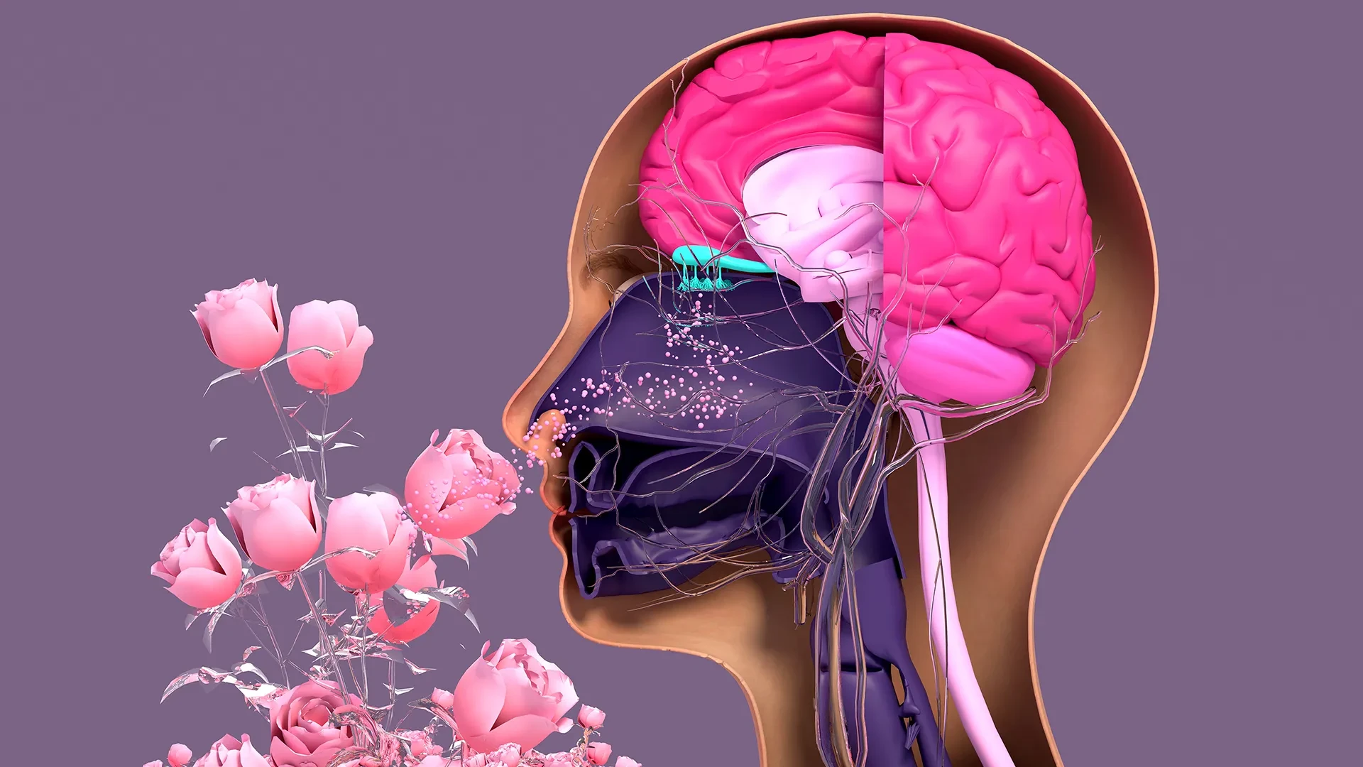
Researchers at Tufts University School of Medicine and the Graduate School of Biomedical Sciences (GSBS) have unveiled a breakthrough in regenerative medicine that could fundamentally change how we understand the human sense of smell. By developing a sophisticated, three-dimensional model to observe nerve tissue regeneration in the nose, the team has discovered that a specific type of stem cell, long assumed to be dormant, actually plays a vital, active role in preserving olfactory function. This discovery challenges established biological models and offers a promising new avenue for treating anosmia—the loss of smell—which has become a major public health concern in the wake of the COVID-19 pandemic.
The human olfactory system is a biological marvel. Unlike the central nervous system, where neurons are notoriously difficult to repair once damaged, the sensory neurons within the nasal cavity possess a unique and remarkable capacity to regenerate throughout an individual’s entire lifespan. This constant renewal is essential, as these cells are frequently exposed to the harsh, unpredictable elements of the external environment, including pathogens, irritants, and toxins.
However, this regenerative process is not infallible. When the system is overwhelmed by severe viral infections—such as those caused by SARS-CoV-2—or subjected to chronic exposure to environmental toxins, the ability of these neurons to replicate and repair themselves can be severely compromised. Aging further compounds this issue, leading to a gradual, often irreversible decline in olfactory sensitivity. For millions of people, this results in a partial or complete loss of smell, a condition that can profoundly impact quality of life, nutritional intake, and emotional well-being.
To better understand the mechanisms behind this failure, the Tufts research team, led by senior author Brian Lin, a research assistant professor in the Department of Developmental, Molecular and Chemical Biology, devised a new, three-dimensional olfactory tissue mouse model. Known as an organoid, this miniature, lab-grown version of nasal tissue provides a controlled environment for scientists to observe exactly how neurons are generated and, crucially, why that process falters during disease or the natural aging process.
The study, recently published in the journal Cell Reports Methods, details how two specific types of stem cells—horizontal basal cells (HBCs) and globose basal cells (GBCs)—interact to build new smell-sensing tissue. Historically, biologists have categorized HBCs as largely dormant, "reserve" cells that only awaken during catastrophic injury. However, the Tufts team’s research presents a more nuanced view of this cellular dynamic.
"Our research suggests that these two stem cells may be interdependent," explains Brian Lin. "One type that we thought was largely dormant—HBCs—may actually play a crucial role in supporting the production of new neurons and the repair of damaged tissue."
By utilizing their novel 3D organoid model, the researchers identified a specific subpopulation of HBCs, distinguished by the expression of the protein KRT5. Contrary to the traditional belief that these cells wait idly for major trauma, the team observed that these KRT5-positive HBCs actively contribute to the daily maintenance and generation of new olfactory neurons. In the organoid environment, these cells acted as essential scaffolds and signaling hubs. When the researchers selectively depleted these KRT5-positive HBCs from the cultures, the organoids’ ability to generate new neurons plummeted, confirming that these cells are fundamental to the regenerative process rather than mere emergency backups.
Beyond the basic mechanics of regeneration, the researchers also sought to understand the impact of aging on these cellular pathways. By growing cells from mice of varying ages within the 3D model, the team identified a clear, age-related decline in regenerative capacity. "We found a decline in the ability of the older mice cells to generate new neurons," Lin notes. "We think this is due to a decrease in the GBC population as we age, but we need to do more work to test this hypothesis and if so, develop ways to rejuvenate them."
A New Tool for Global Research
A significant portion of the credit for the practical utility of this study goes to the lead author, Juliana Gutschow Gameiro. A former Ph.D. student who visited the GSBS from the State University of Londrina in Brazil, Gameiro was driven by a desire to create a tool that was not only scientifically rigorous but also accessible to labs operating with limited funding or specialized equipment.
The need for such a tool has never been greater. As the prevalence of smell loss has surged due to the global COVID-19 pandemic and its association with neurodegenerative conditions like Parkinson’s disease, the scientific community has seen a massive influx of researchers from disparate fields attempting to study olfactory epithelial cells.
"We wanted to develop an easy-to-use model so that non-stem cell biologists and those working in labs with limited resources could use it to better understand how olfactory neurons regenerate and what happens that causes that process to diminish or fail completely," Lin says. By lowering the barrier to entry for this type of research, the team hopes to accelerate the pace of discovery across the global scientific community.
The Path Toward Human Organoids
The ultimate, long-term objective of the Tufts team is to translate the success of their mouse-tissue model into a human organoid system. Such a platform would be a game-changer for pharmaceutical research, providing a reliable, ethical, and efficient way to screen drugs intended to restore the sense of smell in humans.
Currently, pre-clinical research relies heavily on whole-animal testing or existing human cell cultures, both of which have significant limitations. Whole-animal models are expensive, time-consuming, and often fail to perfectly replicate human physiology. Conversely, static cell cultures lack the complex 3D architecture required to mimic the real-world behavior of tissue. While organoids have already been successfully developed for other organs, including the lungs and kidneys, human olfactory tissue has proven particularly difficult to isolate.
The challenge lies in the extraction process. Obtaining pure olfactory tissue from human subjects requires a delicate, invasive procedure where a brush—similar to a COVID-19 nasal swab—is inserted deep into the nasal cavity while the patient is under anesthesia. The problem, as Lin explains, is that the collected sample is a chaotic mixture of cells. "It’s challenging to get pure olfactory tissue from humans," he notes. "Unlike in their mouse model, human respiratory stem cells and olfactory stem cells collected in this process are difficult to separate."
Because the olfactory and respiratory stem cells are often harvested together, researchers have struggled to coax the olfactory-specific cells to grow in a lab environment without being overtaken by the more aggressive respiratory cells. The team’s next major challenge is to refine a technique that is both simple and cost-effective for isolating these specific human olfactory stem cells. Once they successfully conquer this hurdle, the path to testing restorative therapies for those who have lost their sense of smell will be significantly clearer.
By shifting the focus toward the previously overlooked role of HBCs and providing the scientific community with a standardized, accessible 3D model, the researchers at Tufts have laid the groundwork for a new era of olfactory research. As the team continues to refine their techniques, their work offers a glimmer of hope that the mechanisms behind smell loss may soon be fully understood, and potentially, reversed.