In a significant leap forward for olfactory research, scientists at Tufts University School of Medicine and the Graduate School of Biomedical Sciences (GSBS) have unveiled a sophisticated, three-dimensional model designed to study the regeneration of nerve tissue within the nose. This innovative research, recently published in the journal Cell Reports Methods, has overturned long-held assumptions regarding the cellular mechanisms that preserve our sense of smell. Most notably, the team discovered that a specific type of stem cell, previously dismissed by the scientific community as largely dormant, actually serves as a cornerstone for maintaining olfactory function.
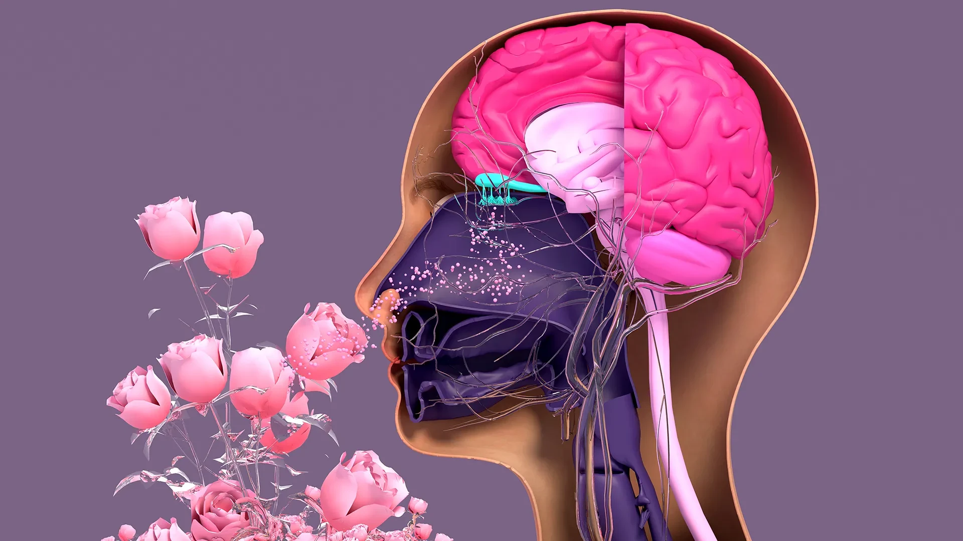
The human sense of smell is a complex biological feat, one that relies on sensory neurons located deep within the nasal cavity. Unlike the rigid architecture of the central nervous system, where damaged neurons often struggle to recover, olfactory sensory neurons possess a remarkable, lifelong capacity for regeneration. This continuous renewal process allows the nose to adapt and repair itself, a necessary defense mechanism given its constant, direct exposure to the volatile environment, pathogens, and environmental toxins.
However, this regenerative capacity is not infinite. A wide array of external and internal stressors—ranging from viral infections, such as COVID-19, to chronic exposure to toxins and the inevitable biological decline associated with aging—can severely impair these cells. When the replication process falters, the result is often a partial or complete loss of smell, a condition that can profoundly diminish an individual’s quality of life and, in some cases, serve as an early indicator of neurodegenerative diseases like Parkinson’s. To better understand why this regenerative system eventually falters, the Tufts research team developed a three-dimensional olfactory tissue mouse model, or organoid. This tool provides a highly controlled environment for scientists to observe how neurons are continually birthed in the nose and to pinpoint the exact moment the process begins to fail.
At the heart of the study is the complex relationship between two specific types of stem cells found in the nasal epithelium: horizontal basal cells (HBCs) and globose basal cells (GBCs). Traditionally, the scientific consensus categorized GBCs as the primary workhorses of neurogenesis, while HBCs were viewed as a dormant "reserve" pool, activated only in the event of severe injury to the nasal lining. The Tufts research suggests a far more nuanced and collaborative reality.
"Our research suggests that these two stem cells may be interdependent," explains Brian Lin, senior author of the study and a research assistant professor in the Department of Developmental, Molecular and Chemical Biology at Tufts. "One type that we thought was largely dormant—HBCs—may actually play a crucial role in supporting the production of new neurons and the repair of damaged tissue."
Through the application of their new organoid model, the team identified a specific subpopulation of HBCs, distinctly marked by the production of the protein KRT5. These cells do not simply sit in waiting; they appear to be active participants in the generation of new olfactory neurons. When the researchers selectively depleted these KRT5-positive HBCs from the organoid cultures, the formation of new neurons was significantly impaired, confirming that these cells are essential players in the regenerative cycle rather than mere bystanders.
The study further investigated the impact of aging on this cellular machinery. By observing cells derived from mice of varying ages, the researchers noted a marked decline in the regenerative capacity of cells taken from older subjects. While the team suspects this decline is linked to a reduction in the GBC population over time, Lin emphasizes that further investigation is required to solidify this hypothesis. "We think this is due to a decrease in the GBC population as we age, but we need to do more work to test this hypothesis and if so, develop ways to rejuvenate them," Lin says.
An Accessible Tool for Global Research
The development of this three-dimensional model was driven by a commitment to scientific accessibility. Juliana Gutschow Gameiro, the lead author of the study and a former Ph.D. student visiting GSBS from the State University of Londrina in Brazil, spearheaded the effort to create a system that is both effective and relatively simple to implement.
The urgency of this work has been amplified by the global pandemic. The high prevalence of smell loss following COVID-19 infections, combined with the established link between olfactory dysfunction and conditions such as Parkinson’s disease, has drawn an influx of researchers from disparate fields into the study of olfactory epithelial cells. Recognizing this, the Tufts team sought to lower the barrier to entry for laboratories that may not specialize in stem cell biology or possess high-end, expensive equipment.
"We wanted to develop an easy-to-use model so that non-stem cell biologists and those working in labs with limited resources could use it to better understand how olfactory neurons regenerate and what happens that causes that process to diminish or fail completely," Lin notes. By democratizing the tools used to study these cells, the researchers hope to accelerate the pace of discovery across the global scientific community.
Toward a Human Organoid and Future Therapies
The ultimate ambition of this project extends beyond the mouse model; the researchers aim to use this framework as a stepping stone toward developing a human olfactory organoid. Such a model would be a transformative tool for drug screening, allowing scientists to test potential therapies for patients suffering from long-term smell loss in a lab setting, rather than relying on animal models or complex, often inefficient human cell cultures.
Organoids—miniature, simplified versions of organs grown in a dish—have already revolutionized research in other fields, with successful models developed for the lungs, kidneys, and other major organs. However, creating a functional human olfactory organoid remains a formidable technical challenge. Obtaining pure, uncontaminated olfactory tissue from human subjects is difficult. The standard collection procedure involves anesthetizing a patient and utilizing a brush—similar to a swab used in a COVID-19 test—to gather cells from deep within the nasal cavity. In humans, unlike in the mouse model, the resulting sample is a mixture of respiratory stem cells and olfactory stem cells, which are notoriously difficult to separate.
For the research team, the path forward is clear: they must now develop a technique that is both cost-effective and precise enough to isolate human olfactory stem cells and encourage them to thrive in a laboratory environment. If successful, this work could provide the foundation for new clinical interventions aimed at restoring the sense of smell for millions of people worldwide. As the team continues to refine their approach, the shift from mouse-based discovery to human-centric therapeutic modeling represents the next major hurdle in understanding the resilience and eventual decline of our most delicate sensory systems.